Boxer Dog Mast Cell Skin Tumors
Overview
Mast cell tumors are seen with the Boxer breed more than any other breed that exists. For this reason, owners should know about these, how to check for them, and what can happen if they appear. In this profile, we will:
- Go over the details of what these are
- Meet a really awesome Boxer dog that had and has several tumors, and endured extensive surgery and recovery
- The importance of prevention and early detection
Please note: AllBoxerInfo is reader-supported. Some of the product suggestions on this page are affiliate links. As an Amazon Associate we earn from qualifying purchases. This is at no extra cost to you and helps keep this site running.
About Mast Cell Tumors
What are mast cells?
Mast cells are found all over the body. They are derived from bone marrow, and are found in all connective tissue, which is tissue that connects, supports or separates other tissues or organs.
They are found in skin (the largest connective tissue on a dog's body - or a human's) and these are also present in the intestines and respiratory tract.
Mast cells serve several purposes:
- Contain chemicals including histamine, heparin, and proteolytic enzymes. These chemicals are activated during an immune system response; such as if a dog is having an allergic reaction or in defense of parasitic attack
- Work to repair tissue
- Play a role in the formation of blood vessels
What are mast cell tumors?
A tumor is any type of abnormal growth.
When mast cell tumors (abnormal growth of a cluster of mast cells) develop on a dog, they can be benign (non-cancerous) or malignant (cancerous). In addition, and importantly, a benign mast cell tumor can spread (metastasize) and become malignant.
There are 3 grades:
Grade 1:
Low potential for metastasis and considered presently benign. They can range in size from small to large. May or may not be removed. This depends on where they appear and if they appear to be growing.
Grade 2:
Potential for metastasis and shows some characteristics of possibly turning malignant. These often extend below the skin and are intricately weaved into subcutaneous tissues, which is the inner most layer of the skin, containing blood vessels and nerves. May or may not be removed; more ahead.
Grade 3:
High potential for metastasis. These are also deep into subcutaneous tissues and have the highest determination to metastasize. Are often removed; however that presents issues as well, more ahead.
Causes
The causes are unknown; however, both genetics and environmental factors are suspected.
Occurrence rate and stats
• Out of all skin tumors found on canines, 20% are mast cell tumors. The Boxer breed has more mast cell tumors that any other breed.
• The age that these appear is, on average, 8 years old. But, they can appear at a much younger age.
• They can spread to lymph nodes, and do in 76% of dogs.
• Occurrence or tumor-related death is 3.5% for the Boxer breed.
• A study by Kansas State University shows that they most often occur on the abdomen or hind leg (40.4%), chest or foreleg (28.9%), genitals or anal region (17.5%) and head or neck (13.2%).
Diagnosing
The vet will take a tissue sample of the tumor by either by Fine needle aspiration or a biopsy and bloodwork will be done.
Treatment for mast cell tumors
Keeping in mind that these tumors can be grade 1 through 3, some may be left but watched carefully, and some may be removed. Removing them presents several challenges:
- When a mast cell tumor is removed, it can trigger large quantities of the chemicals it holds (histamine, heparin, and others) into the body. And this can cause complications including a sometimes dangerous effect on heart rate and blood pressure.
- It is common for the removal site to refuse to heal, leading to open wounds.
- For the surgery to be successful, surrounding tissue must also be removed. It is very difficult to ascertain exactly where the tumor ends and healthy tissue begins. For this reason, a wide portion of healthy tissue is often removed.
There are other possible treatments as well, depending on how aggressive the mast cell tumors are. This includes radiation and chemotherapy.
Prognosis
This mainly depends on the grade of the mast cell tumor and where it is located. In regard to location, prognosis is best for those on the limbs. It is poor for those found on the genitals, muzzle, mouth, and nail bed area of the paws. It is very poor for those found in internal organs or in bone marrow.
In regard to the grade, for grades I and II that were fully
removed, the success rate of no recurrence within 3 years is 90 to 95%.
Meet a Boxer Dog that Had Tumors at a Young Age
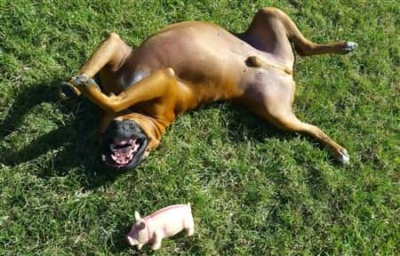
This is Rosco Tiberius Workman
-min(1)-263x350-1920w.jpg)
Rosco is a 4-year-old Boxer dog owned by Janean & Tom Workman of Austin, TX. When he was just 22 months old, Janean discovered a lump on his hind leg. A couple of months later, there was another on his belly and one on his head.
Here, we have Q&A
with Janean, along with photos showing the surgery he endured, the recovery process, and how he is doing now. Please note:
Photos are not for the faint of heart.
Rosco's Story

ABI:
Thanks, Janean, for taking the time to tell us about what happened to your Boxer dog. It's important for other owners to know about this, so they can catch any skin tumors early.
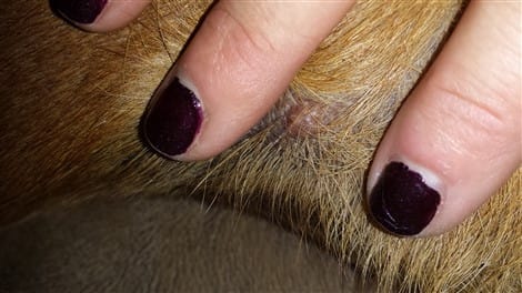
Q: How was it that you discovered the first lump?
A:
I was giving him a massage, and felt the teenie bump.
Q: Rosco had 3 tumors in all?
A:
Yes, I discovered the first one on his leg when he was 22 months old. At about 28 months, the other two were found.
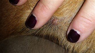
The veterinarian recommended that the one on his leg be removed. We were warned that it would be a hard recovery and a nasty cut.
The other two are very small, and we are keeping an eye on those. The one on his head would involve a very high risk surgery due to its location.
A: Can you tell us about the surgery?
(Again, these photos will not
be for the squeamish)
A:
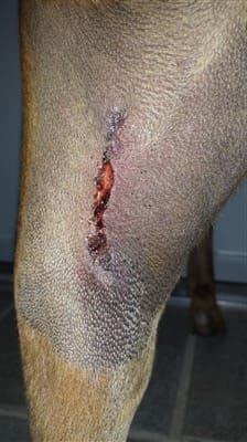
We monitored the growth of the tumor, and when he was 24 months old, the decision was made to remove it.
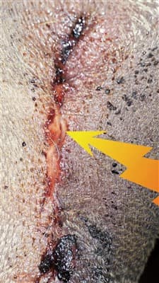
The veterinarian warned us that because of the location, the middle of hamstring muscle, that it would be a deep cut as she would need to make sure 'all of it would be removed'.
He went in for surgery around 9:30 AM, and he was home later that evening.
-min-320x569-1920w.jpg)
When the tumor was removed, it looked like a big bite was taken from his hamstring.
Q: Can you tell us about the recovery process?
Because the Boxer has such a hard time keeping his energy level at low and refrain from jumping, it was almost impossible to keep the stitches from not opening.
The stitches came undone twice, and then he had staples, and then those had to be removed. The total recovery time was about two weeks.
-min-263x350-1920w.jpg)
Having a Boxer that cannot exercise for 2 weeks is nearly impossible to bear. The crying of wanting to go for a walk or a hike, and not being able to communicate the healing process was very stressful.
Thank goodness the ones on his belly and behind his left hear have remained smaller.
Q: How is Rosco's mobility now? Does he have any after effects from this? How is his ability to run, jump, etc.?
A:
He loves to jump and he has what we call the 'Zoomies', when he drops his bottom end and just sprints around, then freezes, then sprints around again. It's my favorite silly move ever!
-min-450x238-1920w.jpg)
He's got the biggest heart in the world, UBER spoiled, and loved beyond belief.

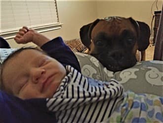
He loves his Mommy (me) and Daddy (my husband) and his new baby brother Wyatt, my 1st born son - only 2 months old now. Photo: Rosco watching over 10 day old Wyatt.
ABI:
A huge thank you to Janean for sharing this story of her Boxer dog Rosco (while taking care of a newborn baby!) and for supplying the photos to show us what this awesome Boxer dog persevered through.
This is a wake-up call for all Boxer owners who do not take steps to help prevent and check for tumors. So, we'll dive into that next.
You can also opt for a wipe, like Petkin Canine Sunwipes , which can be an easy method to apply as well, and is SPF 15.
2. Have your Boxer eat clean.
Your Boxer puppy or dog should not be ingesting any chemicals.
And some foods are packed
with them. You've really got to watch out for artificial chemical preservatives, coloring, and flavoring. These are nasty; they can cause skin reactions, digestive upset, and
long-term ingestion of synthetic food additives are linked to cancer.
If you aren't positive that your Boxer is eating super healthy, it'll be time to reassess what you're giving him. One of our newest recommended foods is Wellness CORE Natural Grain-Free
 ; this is a 5-star food that is hard to beat.
There's great options including Ocean Whitefish, Herring & Salmon, Turkey & Chicken, and Wild Duck, Turkey, Boar & Rabbit. They also have a reduced fat formula.
It has ONLY all natural ingredients. There is zero wheat, corn, soy, or meat by-products. It has no artificial colors, flavors or chemical preservatives. And it's made in the USA.
3. Don't let your Boxer drink straight tap water.
If you do, this can be slowly poisoning your Boxer dog. It came out not too long ago that Chromium-6 was found in the tap water of 31 cities and affecting over 200 million people; this is a known carcinogen (you might remember this from the Erin Brockovich movie).
A suspected carcinogen, 1,4-dioxane, was found in Long Island's water supply.
Everything from rock fuel, to pesticides, to arsenic can be in tap water. Typically, in any given year there are 100 chemicals in drinking water that are unregulated by the FDA.
But, just as worrisome is what is known and legally allowed to be in it. Just one of the hundreds of concerns is hexavalent chromium which is linked to cancer and gastrointestinal tumors.
So, while it can't
get it all,
connect a water filtering system to your kitchen sink, use a water filtering pitcher like the pH RESTORE Water Pitcher Ionizer By Invigorated Water , or though it's not the most cost effective, go with gallons of spring water only.
4. Early Detection.
It's easy, but far too overlooked by many owners:
Check your Boxer. Run your hands over every area of his body to feel for lumps.
It's suggested to do this every week. If you find any sort of lump, bump, or growth, immediately call the vet and get him to the clinic to be examined. The earlier a tumor is found and diagnosed, the better the prognosis.
Set a reminder on your phone, or make it a weekly ritual (like a half hour before your favorite show, etc.) so you won't forget.